Dr. Stephanie Frankel’s
Case of the Week: Myelinated Retinal Nerve Fiber Layer (MNFL)
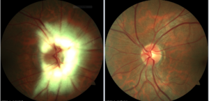
52-year-old male presents to clinic for evaluation of a bump on his right upper lid, which has existed for about two weeks. He has no significant ocular history. His medical history is positive for hypertension which is controlled with Enalapril. Best corrected visual acuity is 20/20 OD and 20/25 OS. His intraocular pressures were 18 OU. Slit lamp examination revealed a chalazion of the lateral right upper lid which going to be managed with excision at follow up. Posterior examination revealed the images below.

What is your diagnoses and management?
This condition is known as Myelinated Retinal Nerve Fiber layer of the Optic nerve (MNFL). This occurs when there is abnormal myelination of the nerve fiber layer anterior to the lamina cribosa. It presents as striated patches with feathery borders in the superficial retina. This occurs in about 0.5-1% of the population. Most patients are symptomatic; however visual function can be affected in few cases. It is usually present at birth and continues to remain static majority of the time. Generally, these lesions do not require any treatment, however, it is recommended that you consider formal visual field testing to rule out concomitant neuro-ophthalmic issues as well as assessing impact the lesion may have on visual function.
To schedule your eye exam with Dr. Frankel, give our office a call or simply schedule an appointment via ZocDoc.
